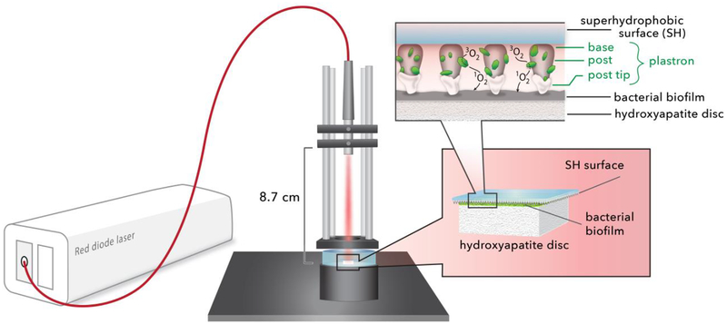
Figure 1.
A schematic of the superhydrophobic device: a red diode laser (669 nm) is coupled to an optical fiber; the output SMA ferrule is mounted such that the light is directed downward. The superhydrophobic (SH) surface, printed on a 130 μm thick coverslip, is placed tip-face down on the bacterial biofilm. SiO2 nanoparticles are used to cap the SH surface. Singlet oxygen traverses the plastron to reach the biofilm, where inactivation then takes place.

