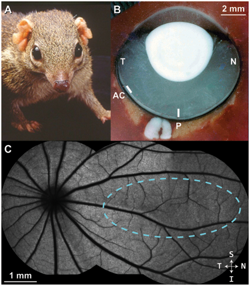
Figure 1 –

Tree shrew ocular anatomy overview. (A) A northern tree shrew showing laterally-placed eyes and binocular visual field. (B) Horizontal section through a flash-frozen tree shrew right eye. N = nasal; T = temporal; P = approximate posterior pole; AC = approximate location of area centralis. In contrast to primates, the optic disc is located in the temporal retina; the posterior pole region is nasal to the optic disc. (C) A montage of blue autofluorescence images in the tree shrew retina indicating the vascular pattern radiating outward from the optic disc. In this study, cardinal axes (superior/inferior, nasal/temporal) were defined relative to the ONH, as it was the most distinct retinal feature. Dashed cyan outline = approximate visual streak (Müller and Peichl, 1989).
