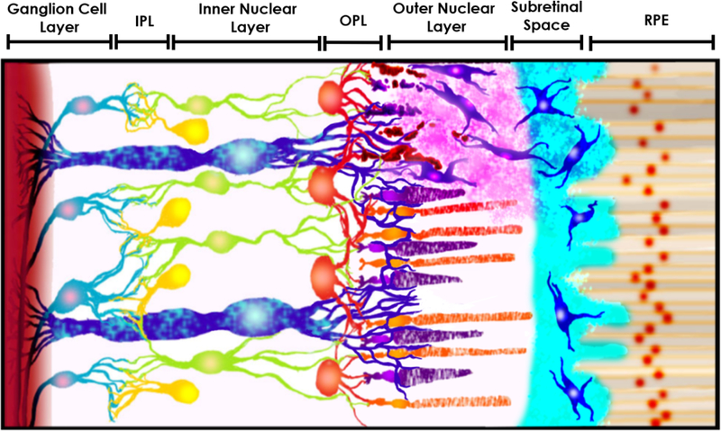
Figure 1.

Schematic of transplanted retinal progenitor cells (RPCs) migrating into damaged host retinal tissue. The schematic provides a representative cross-section of retinal tissue from the retinal pigment epithelium (RPE) at the eye posterior to the ganglion cells of the optic nerve (from right to left, not to scale). RPCs (shown in blue) are transplanted in the sub-retinal space between the RPE and outer nuclear layer (ONL), in which the native rod and cone photoreceptors reside. Transplanted RPCs then migrate into retinal tissue to synaptically integrate in the outer plexiform layer (OPL) with native horizontal cells (HCs), bipolar cells (BCs) in the inner nuclear layer (INL) in turn with ganglion cells (GCs) and amacrine cells (ACs) in the inner plexiform layer (IPL) to restore vision. The figure also features the presence of Müller glia cells (MGCs) along all three main retinal layers. Modified after Thakur et al 2018.
