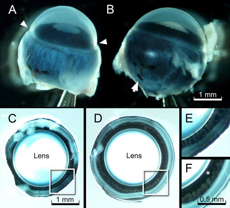
Figure 2.
Effect of pressurized fixation on circumlental space. A. Eye of a one-month-old mouse after conventional immersion fixation. Note the constriction at the limbus (arrowheads). B. Appearance of the contralateral globe, fixed under an internal pressure of 27 cm H20, as described in the text. Note that the eye retains its smooth, spherical contours. The puncture wound caused by the fixation needle is visible in the posterior sclera (arrow). C. The deflated eye (from A) photographed from the posterior aspect, after removal of the sclera. D. The appearance of the eye (from B) fixed under pressure. E and F. Higher magnification views of the boxed regions from C and D, respectively. Note that the circumlental space (*) collapses in the eye prepared using conventional immersion fixation (E) but remains patent in the pressure-fixed eye (F).

