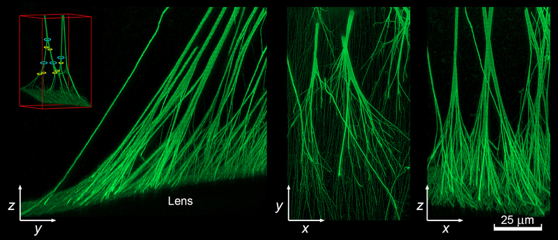
Figure 6.
High resolution reconstruction of the attachment of the posterior zonular fibers to the lens surface visualized in yz, xy, and xz projections. Fibers were visualized using anti-LTBP2 immunofluorescence. The image stack was deconvolved before rendering. Note that the fibers branch multiple times as they approach the lens. The inset shows an example of the trunks (blue) and branches (yellow) on either side of a nodal point (see text for details).

