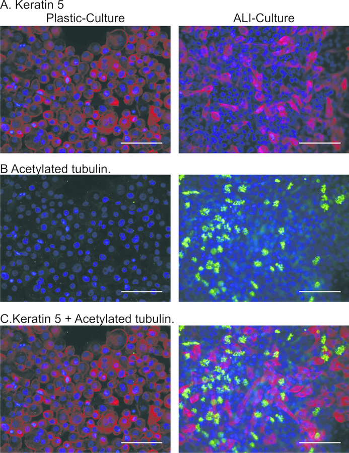
Figure 4. Characterization of airway epithelial cell marker protein expression by immunostaining.
Expression of KRT5 (red; basal cells) and acetylated tubulin (green; ciliated cells) were assessed in PHLE by immunocytochemistry. Counter-stain is DAPI (blue). During non-ALI culture, a majority of cells express KRT5 but not acetylated tubulin. Upon differentiation at ALI, the number of KRT5 expressing cells decreases, and acetylated tubulin staining appears in cells not expressing KRT5. Scale bar is 100 μm.

