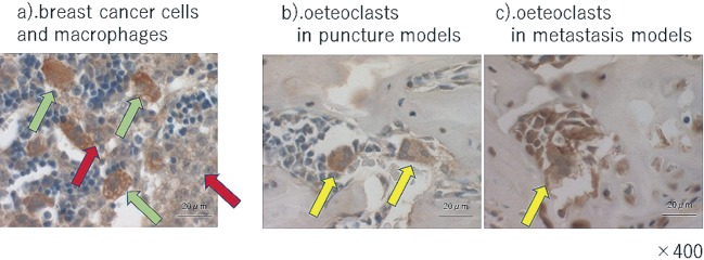
Figure 4.
Immunohistochemistry of IL-6 a) breast cancer cells. Red arrow heads indicate the breast cancer cells and green arrowheads shows macrophages surrounding the breast cancer cells. They were IL-6-positive. b).and c). osteoclasts in puncture models and metastasis models. Arrow heads indicate the osteoclasts. The osteoclasts in the puncture models and metastasis models are both IL- 6 positive. The scale bars are 20μm.

