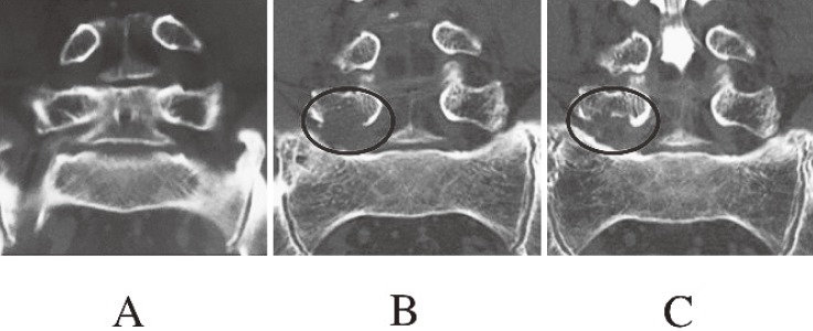
Figure 5.
Coronal images of reconstructed three-dimensional computed tomography in patient 4.
A. Before surgery.
B. Six months after surgery. Clearance under the L5 pedicle was enlarged by removing the caudal aspect of the L5 pedicle (circle).
C. Thirty months after surgery. Clearance under the L5 pedicle decreased because of bone regrowth of the L5 pedicle (circle).

