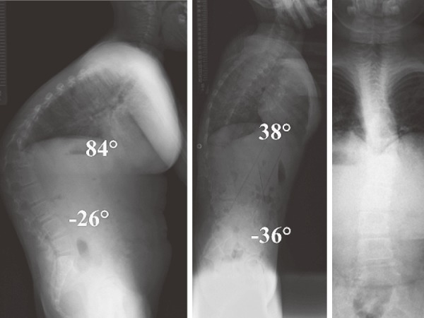
Figure 2.

Preoperative radiograph. X-ray imaging showed kyphosis of 84° between T6 and L3 with T12 as the apical vertebra and kyphosis was corrected to 38° under traction. Local kyphosis was 51° between T11 and L1 with slight scoliosis in anteroposterior view.
