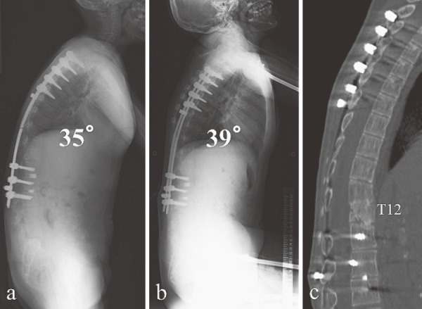
Figure 4.

(a) Postoperative radiograph. Kyphosis was corrected from 84° to 35° between T6 and L3. Local kyphosis improved from 51° to 16°. Lumbar lordosis decreased from 26° to 6°. (b) Final follow-up radiograph (6 years after surgery). The kyphosis was maintained at 39° without vertebral fracture at the latest follow-up. (c) Final follow-up computed tomography (6 years after surgery). T12 pedicle subtraction osteotomy was performed, and the thoracolumbar junction became flat.
