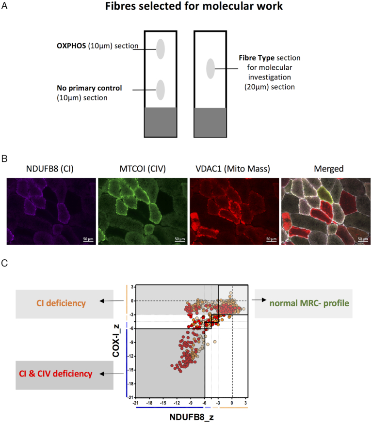
Figure 1.
Quadruple immunofluorescence and selection of fibres for the molecular analysis of six patients. (A) Three serial muscle sections were taken from each patient biopsy for OXPHOS and fibre type analysis. The OXPHOS section was sequentially overlaid with all primary and secondary antibodies, whereas the no primary control section was only overlaid with the laminin antibody and all secondary antibodies. The fibre type section was overlaid with antibodies, detecting type I and type II myosin heavy chain. (B) Example quadruple immunofluorescent images for patient 3 with a TWNK variant. NDUFB8 is used as a complex I (CI) marker, MTCOI (COXI) as a complex IV (CIV) and VDAC1 as a mitochondrial mass marker. The merged image also includes the membrane marker laminin; scale bar: 50 μm. (C) Each cell of the fibre typed section was then matched with the corresponding fibre from the quadruple immunofluorescence staining. Depending of the ODporin values and the different fibre types (type 1, type 2a, type 2x), fibres were selected randomly out of each group for molecular work.

