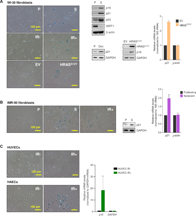
Figure 1.
Senescence models. (A) The phenotype of proliferating WI-38 fibroblasts rendered senescent by extended culture [P (proliferating) cells at PDL25; S (senescent) cells at PDL50-59], exposure to ionizing radiation [IR-, proliferating; IR+, senescent by 10 days after exposure to 10 Gy] or expression of HRASG12V to trigger oncogene-induced senescence (OIS) [EV, proliferating cells expressing empty vector; HRASG12V cells expressing oncogenic RAS, 5 days after infection and selection with puromycin] was studied by assessing senescence-associated (SA)βGal activity (micrographs), by western blot analysis of senescence marker proteins showing higher (p16, p21, p53) or lower (SIRT1) levels with senescence (middle) or by RT-qPCR analysis of p21 mRNA levels (graph). WI-38 fibroblasts at PDL25 were treated with Doxorubicin (2 μg/ml for 24 h and harvested 5 days later; senescence was assessed by western blot analysis of p21 expression levels. (B) The phenotype of IMR-90 cells that were either proliferating (P) or were rendered senescent by replicative exhaustion (S) or exposure to IR (IR+) was assessed as explained in panel (A). (C) The senescent phenotype of proliferating (IR-) and senescent (IR+) HUVECs and HAECs (4 Gy, 10 days after exposure) was assessed by monitoring SA-βGal activity (micrographs) and p16 mRNA levels (graph). Data in graphs (A–C) represent the means ± S.E.M. from three independent experiments.

