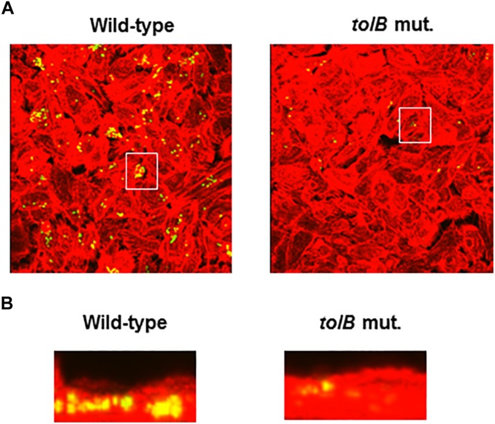
FIGURE 3.
Fluorescent images of aggregated UPEC cells within bladder epithelial cells (HTB-9). Bacteria carrying a Green fluorescence protein (GFP) expression plasmid, pTurboGFP-B and HTB-9 stained with rhodamine- phalloidin were imaged as, respectively, green and red fluorescent colors on the microscopy using 60× objective. Overlooking images (A) and cross sectional images in the white box indicated in panel A (B) were acquired for each sample. The experiment was repeated twice and similar results were obtained.

