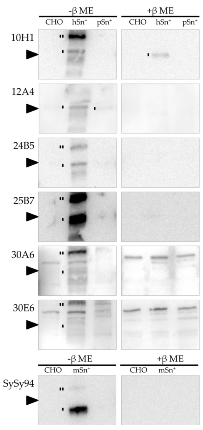
Figure 3.
Western blot analysis of the different mAbs. Lysates of CHO, hSn+-CHO, pSn+-CHO, and mSn+-CHO were run under non-reducing (-β mercaptoethanol (ME)) and reducing (+β ME) conditions. The newly developed mAbs were added to the blots followed by an anti-mouse or anti-rat horseradish peroxidase (HRP)-labeled secondary antibody. Sn monomers and dimers are indicated with one line and two lines respectively. The arrow indicates 220 kDa.

