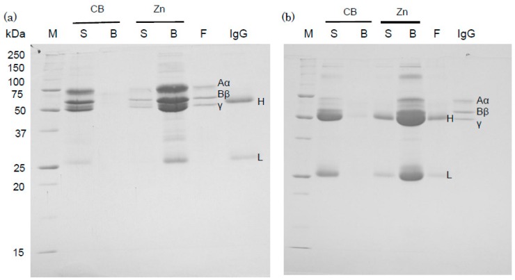
Figure 4.
Binding of human IgG and fibrinogen to Zn-beads which were saturated with fibrinogen and IgG, respectively. After binding of human fibrinogen (600 µg) or IgG (500 µg) to Zn-beads or CB (net volume of beads per sample: 20 µL each), aliquots (20 µL) of human IgG (100 µg) or fibrinogen (100 µg) in PBS were added to protein-saturated Zn-beads and incubated with: fibrinogen in (a) and IgG in (b). As described in the “Experimental Section”, the supernatant (S) and bead samples (B) obtained after centrifugation were subjected to SDS-PAGE. Human fibrinogen (F) and IgG samples were also applied (2 µg/lane). Their separated subunit bands derive from fibrinogen (Aα, Bβ and γ) and IgG (H and L). M represents marker proteins.

