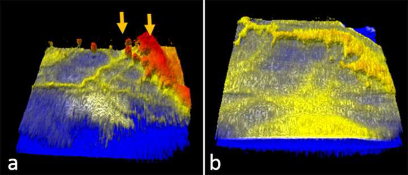
Figure 3:

Representative optical coherence tomography colorized three-dimensional (3D) volumes of a preterm infant with zone I stage 3 retinopathy of prematurity, prior to (a) and three weeks after (b) intravitreal bevacizumab injection. The yellow arrows in a indicating extraretinal neovascular tissue is seen regressing three weeks later.
