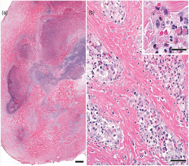
Figure 3.
Photomicrographs of an excisional biopsy specimen of one of the sublumbar masses in the cat. (a) The well-circumscribed mass consists of sclerotic fibrous tissue with central necrosis. Hematoxylin and eosin stain; bar = 1 mm. (b) Hypertrophied fibroblasts are in the sclerotic tissue and in the scattered foci of eosinophilic inflammation. Hematoxylin and eosin stain; bar = 60 μm. (inset) Higher magnification of an aggregate of eosinophils mixed with other leukocytes and fibroblasts. Hematoxylin and eosin stain; bar = 25 μm

