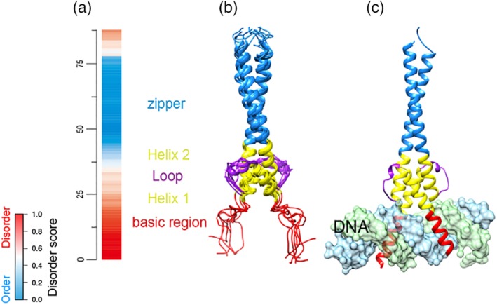
Figure 5.

DNA‐induced folding of the disordered basic region from HLH domain of human Max protein. (a) VSL2b disorder prediction for the sequences. Red: disorder; blue: order; white: ambiguous. (b) NMR structure of the HLH domain in the absence of DNA (PDB id: 1R05). Basic region: 1–18 residues. Helix–loop–helix: 19–53 residues. Leucine zipper: 54–87 residues. (c) X‐ray structure of the HLH domain bound with DNA (PDB id: 1NKP). The two structures represent the same regions (23–102 residues) from human Max protein (UniProt ID: P61244). DNA chains are colored in light blue and green. NMR, nuclear magnetic resonance; PDB, PDB, Protein Data Bank; VSL2B, disorder predictor trained on V = variously characterized proteins with S = short and/or L = long IDRs, version 2b
