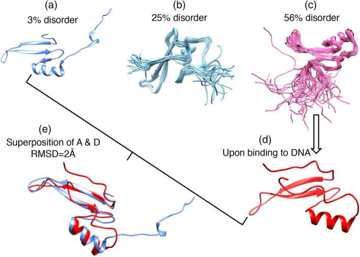
Figure 6.

Diversity of structure/disorder in the MBD domain (PF01429). Top: multiple sequence alignment was carried out by T‐coffee and displayed by ESPript 3.0. Predicted disorder for the sequences increases from top (structure) to bottom (full disorder). The asterisk (*) at the bottom of the alignment indicates highly conserved DNA‐binding sites four available structures were shown below. (a) A structural member (PDB id: 3VXV, chain A). (b) A mediate disordered member (PDB id: 2MOE, chain A). (c) A higher disordered member (PDB id: 1QK9, chain A). (d) A fully disordered member in its DNA bound state (PDB id: 6CCG, chain A). (e) Superposition of the least and fully disordered member (sequence identity 42%) in their bound state. The binding partners are not shown here. MBD, methly‐CpG binding domain; PDB, PDB, Protein Data Bank
