
Figure 1.
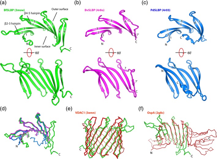
Crystal structures of SLBPs. (a) BfSLBP (green), (b) BvSLBP (magenta), and (c) PdSLBP (blue). The strands are labeled sequentially from 1 to 9. β‐hairpins (βx‐y, where x and y are two adjacent β‐strands forming a hairpin, e.g., β2‐3 hairpin) are referred to as up hairpins (i.e., the loop is on the top, as depicted), or down hairpins (i.e., the loop is at the bottom, as depicted). (d) Superimposition of BfSLBP (green), BvSLBP (magenta), and PdSLBP (blue). (e) Structural comparison between BfSLBP and VDAC1 porin (PDB ID 3emn), and (f) between BfSLBP and OspA (PDB ID 2g8c). BfSLBP is colored green, and the other molecule is red.