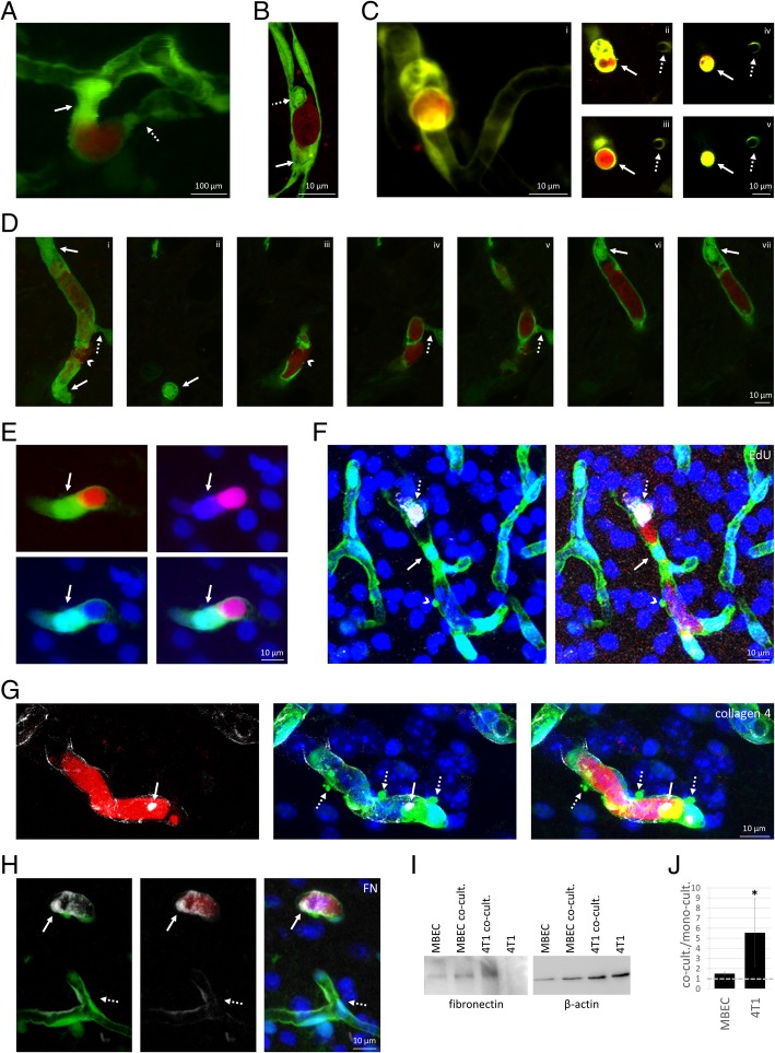
Fig. 2.
Vascular changes induced by metastatic cells before extravasation into the brain parenchyma. a: Two-photon micrograph (z-projection) of an endothelial plug (arrow) and vascular constriction (dotted arrow) in the neighbourhood of an arrested breast cancer cell, on day 5 after tumour cell injection. b: Confocal z-projection of endothelial plugs completely (arrow) or partially (dotted arrow) obstructing the cerebral capillary up- and downstream of an arrested metastatic cell (day 4 after inoculation). c: Complete obstruction of the vessel lumen by the tumour cell-induced endothelial plug in 3D (i) and cross-section (ii-v) two-photon microscopy images (day 5). Arrow indicates tumour cell-induced plug formation in different optical slices. Dotted arrow points to a capillary not affected by cancer cells. d: 3D representation (i) and z-sections (ii-vii) of endothelial plugs (arrows) and vasoconstriction (dotted arrow) closing all capillary branches in the proximity of a transmigrating tumour cell (arrowhead) (confocal micrograph, day 5). e: Confocal z-projection indicating the endothelial nucleus in the plug (arrow) obstructing the capillary next to the cancer cell. f: EdU-positivity (grey) of intraluminal tumour cell, but not of endothelial nucleus in the plug (confocal z-projection). g: Up-regulation of collagen secretion (arrow, grey) in the neighbourhood of arrested carcinoma cells (confocal z-projection). Dashed arrows indicate endothelial blebs. h: Up-regulation of fibronectin secretion (FN, grey) in the neighbourhood of arrested carcinoma cells (confocal z-projection, arrows). Dashed arrows indicate basal fibronectin expression in non-affected capillaries. i: Representative western-blot image of fibronectin expression in mouse brain endothelial (MBEC) and tdTomato-4T1 cells in mono- and co-culture. B-actin was used as loading control. j: Fibronectin protein levels normalized to β-actin in co-cultured vs. mono-cultured MBECs and tdTomato-4T1 cells (average ± SD) (western-blot quantification). N = 3 independent experiments. * P < 0.05 (t-test). Red = tumour cells (tdTomato), green = endothelium (YFP), blue = nuclei (Hoechst)

