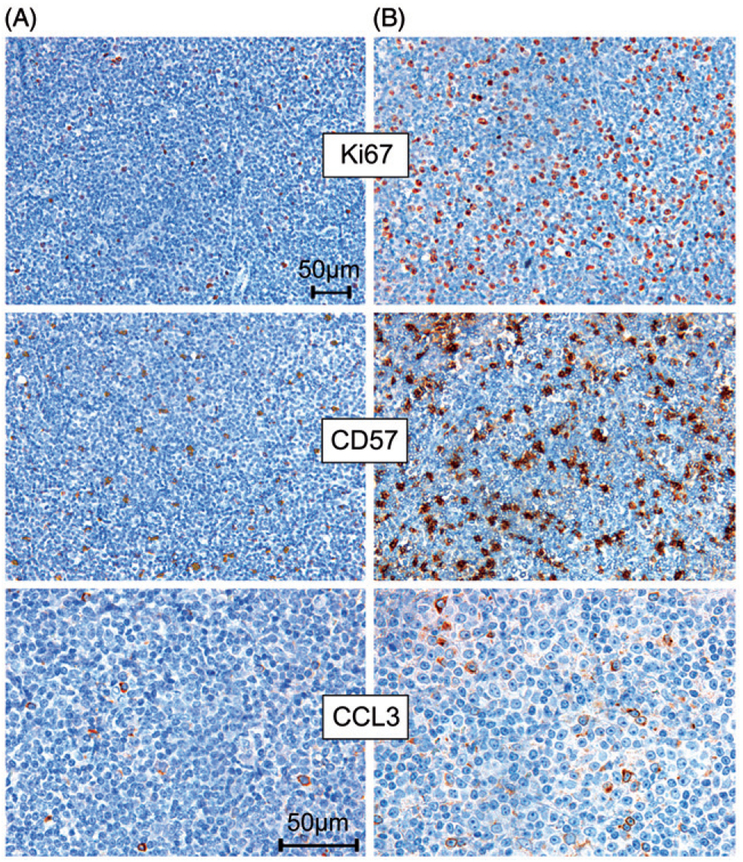
Figure 3.
Increased CCL3 protein expression and a higher number of CD57 positive cells in a case with prolymphocytic progression and increased proliferation (measured by Ki-67 immunohistochemistry) (B), compared with an earlier lymph node excisional biopsy from the same patient with lower proliferation (A). Pictures of Ki-67 and CD57 immunostains were originally photographed at 200× magnification and pictures of CCL3 immunohistochemical staining were originally photographed at 400× magnification.

