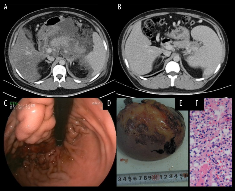
Figure 2.
A 30-year-old male patient was diagnosed with severe acute pancreatitis associated with hyperlipidemia. (A) A contrast-enhanced computed tomography (CT) scan was performed at one week after onset. The pancreas was surrounded by diffuse inflammation. (B) Repeat CT was performed one year later and an enlarged spleen was present. (C) The varices at the fundus of the stomach were complicated by bleeding as shown on endoscopy. (D) Splenectomy and pericardial devascularization were performed due to severe upper gastrointestinal bleeding. (E) Photomicrograph of the histology of the spleen shows congestion with atrophy and expansion of splenic sinus. Hematoxylin and eosin (H&E).

