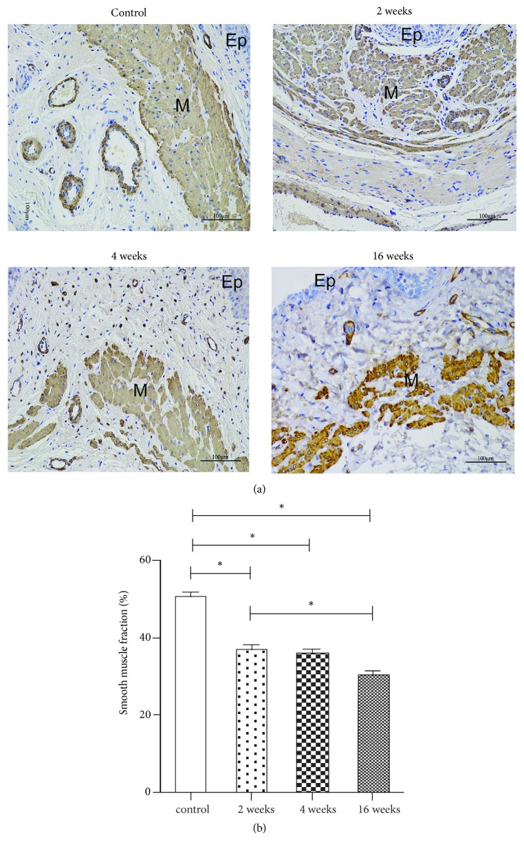
Figure 3.
Morphology and quantity of the nonvascular smooth muscle content in the muscularis of the proximal vaginal wall. (a) Immunohistochemistry images (×200) of a-SMA: Group 1 (control group), Group 2 (2 weeks after ovariectomy), Group 3 (4 weeks after ovariectomy), and Group 4 (16 weeks after ovariectomy). The fractional area of smooth muscle in the muscularis in the four groups (b). The data are presented as the mean±SEM of n=6 animals/group. Scale bar represents 100 μm. Significant differences (P<0.05) are denoted (∗). Ep: epithelium, M: muscularis.

