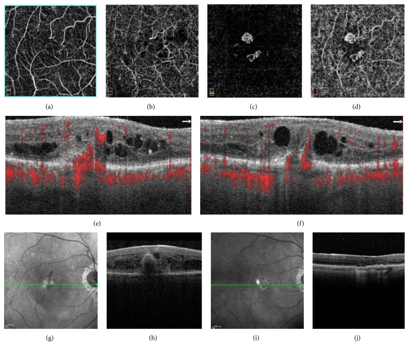
Figure 2.
(a)-(d) Optical coherence tomography angiography images (6 × 6 mm) revealed the progression of the tuft-shaped lesion from the superficial layer to choroid capillary. (a) Superficial capillary plexus segmentation. (b) Deep capillary plexus segmentation. (c) Outer retinal layer segmentation. (d) Choriocapillaris segmentation. (e)-(f) B-Scans optical coherence tomography angiography showed clearly two distinct intraretinal neovascularizations originated in the superficial layer descending into a tuft-shaped lesion toward the sub-RPE space, respectively above (e) and under (f) the fovea. (g)–(h) Spectral Domain-Optical Coherence Tomography at baseline. (i)–(j) Spectral Domain-Optical Coherence Tomography after treatment.

