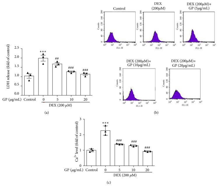
Figure 2.
GP decreases DEX-induced LDH release and Ca2+ concentration increase in C2C12 myotubes. (a) After treatment with DEX for 24 h, cells were incubated with GP (5–20 μg/mL) for 24 h. LDH release was measured by spectrophotometry. (b) After treatment with DEX for 24 h, cells were incubated with GP (5–20 μg/mL) for 24 h. Ca2+ concentration was measured by flow cytometry. (c) Histogram analysis of Ca2+ concentration. Data are expressed as the mean ± SD (n = 3); ∗∗∗p < 0.001 compared to the control group; ## p < 0.01 and ### p < 0.001 compared to the DEX group.

