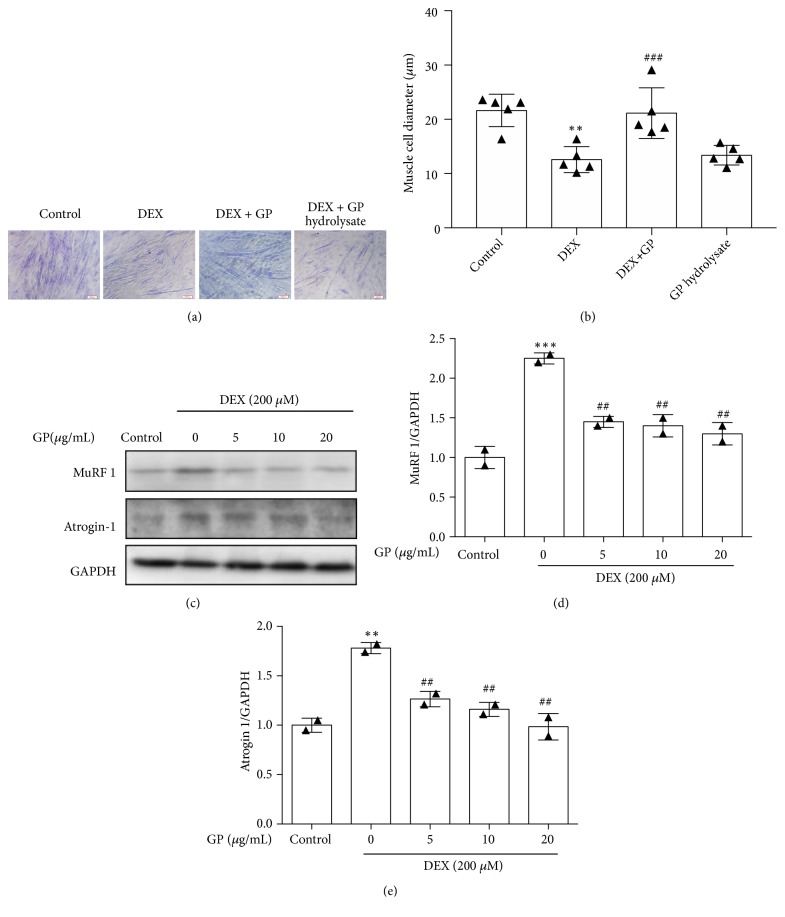
Figure 3.
GP reduces DEX-induced C2C12 myotube atrophy. (a) Representative photographs of C2C12 myotubes for the control, DEX, DEX+GP hydrolysate treatments. (b) Comparison of the cell diameters among the four groups measured after completion of the experiments. (c) After treatment with GP for 24 h, the levels of MuRF1 and atrogin1 in DEX-injured C2C12 myotubes were detected by Western blot analysis. (d) The relative expression of MuRF1 was quantified by densitometry analyses. (e) The relative expression of atrogin-1 was quantified by densitometry analyses. GAPDH was used as the loading control. Data are expressed as the mean ± SD (n = 3); ∗∗p < 0.01 and ∗∗∗p < 0.001 compared to the control group; ##p < 0.01 and ###p<0.001 compared to the DEX group.

