Figure 3:
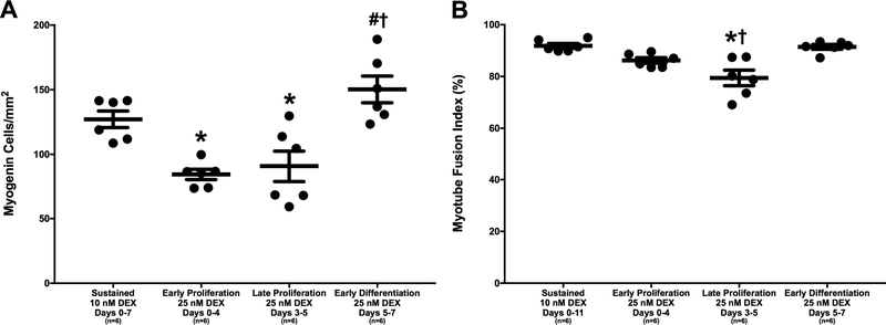
DEX Effects on Myoblast Differentiation into Myotubes. (A) Myogenin-positive cell density, indicating fusion of myoblasts into myotubes, observed with myogenin immunostaining on day 7. Myogenin-positive cell density was significantly lower with administration of 25 nM at early and late stages of proliferation compared to sustained administration of 10 nM DEX and administration of 25 nM DEX at early differentiation (p=0.012, p=0.036, p<0.001, and p<0.001, respectively). (B) Myotube fusion index, representing the percentage of myonuclei associated with a myotube containing 4 or more myonuclei, determined from α-actinin and DAPI staining on day 11. Post hoc analysis indicates a significantly lower myotube fusion index with administration of 25 nM DEX at late proliferation compared to both sustained administration of 10 nM DEX and administration of 25 nM DEX at early differentiation (p<0.001 for both). Thick bars show the mean; error bars indicate standard error. *Indicates a significant difference from sustained 10 nM DEX, †from late proliferation 25 nM DEX.

