Figure 4:
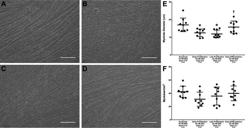
DEX Effects on Myotube Size and Density. Light microscopy images of monolayers were observed before monolayer delamination on day 14. Representative images are shown above for (A) sustained administration of 10 nM DEX (days 0–14), administration of 25 nM DEX at (B) early proliferation (days 0–4), (C) late proliferation (days 3–5), and (D) early differentiation (days 5–7). All images were analyzed for (E) myotube diameter and (F) myotube density. ImageJ analysis indicates that the average myotube diameter is significantly lower with administration of early and late proliferation 25 nM DEX compared to sustained 10 nM DEX (p=0.015 and p=0.005, respectively). Comparison of 25 nM DEX-receiving groups indicates that the average myotube diameter produced through the administration of 25 nM DEX at late proliferation was significantly lower than that produced with administration of 25 nM DEX at early differentiation (p=0.045). There were no significant differences found between the myotube density of the four groups. Scale bars = 100 μm. Thick bars show the mean; error bars indicate standard error. *Indicates a significant difference from sustained 10 nM DEX, †from late proliferation 25 nM DEX.

