Figure 5:
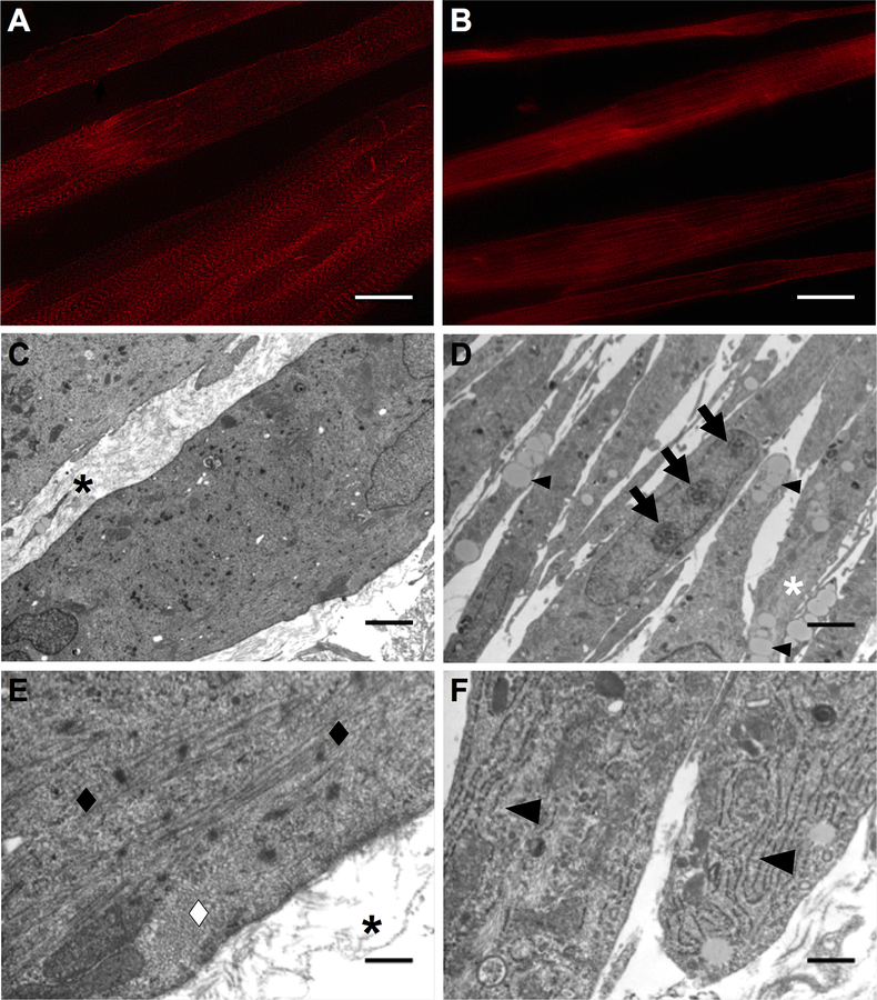
DEX Effects on SMU Structural Maturation. Representative images of α-actinin immunostaining on day 11 following (A) sustained administration of 10 nM DEX (days 0–11) and (B) administration of 25 nM DEX at early differentiation (days 5–7). The greatest advancement in sarcomeric structure and alignment of muscle fibers was observed with sustained administration of 10 nM DEX. Transmission electron micrographs of longitudinal sections of SMUs following (C, E) sustained administration of 10 nM DEX (days 0–16) and (D, F) administration of 25 nM DEX at early differentiation (days 5–7). Sustained administration of 10 nM DEX images show larger myofibers with an abundance of collagen ECM between myofibers, indicated by black asterisks in (C, E), while administration of 25 nM DEX at early differentiation images show smaller myofibers and the absence of collagen ECM between myofibers. Black and white diamonds in (E) indicate sarcomeres in longitudinal and cross orientation along the length of the myofibers observed with sustained administration of 10 nM DEX, respectively. No sarcomeres were observed with administration of 25 nM DEX at early differentiation, but TEM images showed an abundance of myofilaments, lipids droplets, and nucleoli in myonuclei, indicated by a white asterisk, small arrowheads, and arrows in (D), respectively. In addition, large arrowheads in (F) indicate an increase in rough endoplasmic reticulum in the cytoplasm of myocytes with administration of 25 nM DEX at early differentiation. Scale bars in (A, B) 20 μm, (C, D) 2 μm, (E, F) 400 nm.

