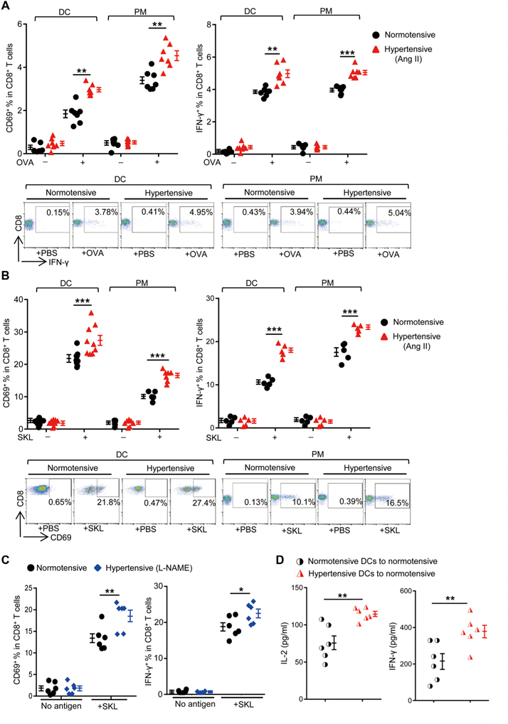
Fig. 3. APCs from hypertensive mice present antigens more efficiently.

(A and B) Splenic DCs and peritoneal macrophages (PM) from normotensive or Ang Il-induced hypertensive C57BL/6 mice were pulsed with 10 μM OVA (A) or 50 pM SIINFEKL peptide (SKL) (B) in vitro and were then coincubated with OT-I T cells. The percentages of CD69+ (after 4 hours) or IFN-γ+ cells (after 6 hours in the presence of brefeldin A) among OT-I T cells were measured by flow cytometry. Representative flow cytometry dot plots of IFN-γ and CD69 expression are shown. (C) Splenic DCs from normotensive or L-NAME-induced hypertensive mice were pulsed with SIINFEKL and then coincubated with OT-I T cells. The percentages of CD69+ or IFN-γ+ cells among all CD8+ T cells were measured. (D) Splenic DCs from normotensive or Ang II-induced hypertensive mice were loaded with SIINFEKL for 4 hours ex vivo. Cells were then transferred intravenously into naїve C57BL/6 mice. Seven days later, splenocytes from the recipients were restimulated with SIINFEKL and the secretion of IL-2 and IFN-γ was measured. Data are means ± SEM. *P < 0.05; **P < 0.01; ***P < 0.005.
