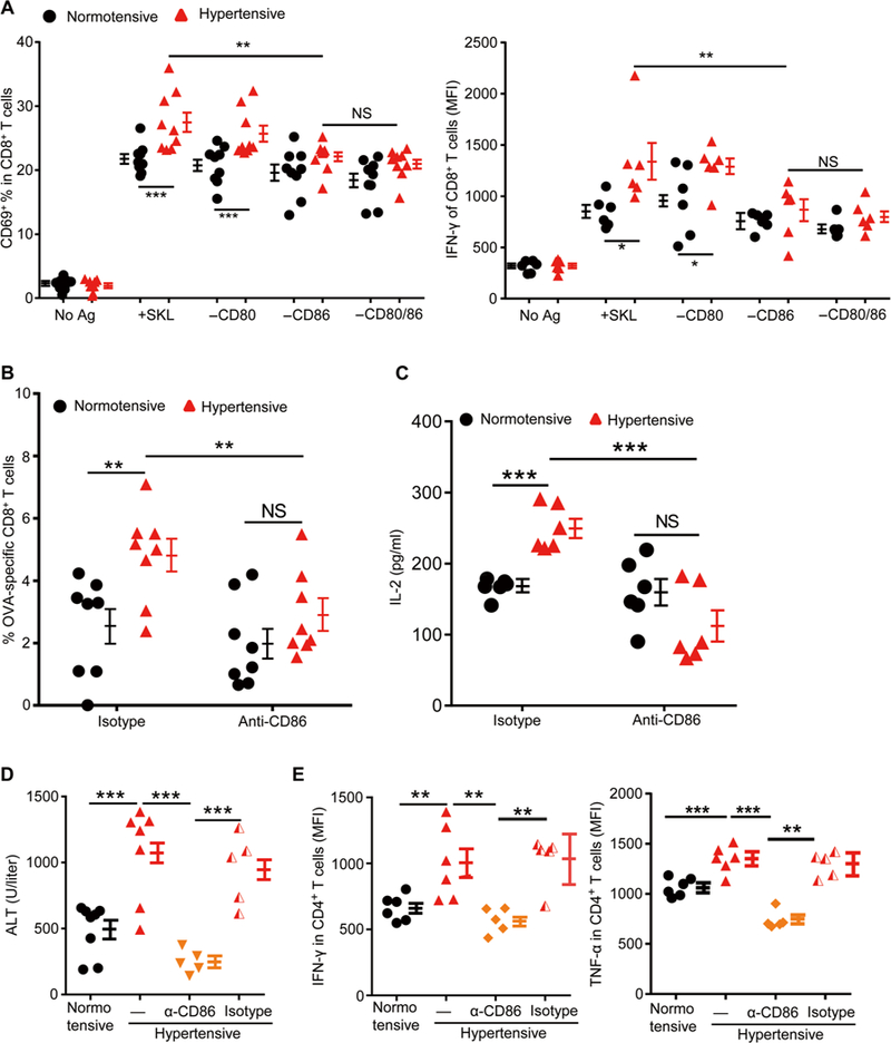
Fig. 5. Blocking CD86 eliminates the overactivation of immune responses in hypertensive mice.

C57BL/6 mice were sham-treated (normotensive) or Ang Il-treated (hypertensive) for 2 weeks. (A) Splenic DCs were purified. Some of the cells were pulsed with SIINFEKL peptide. Some cells were also treated with an anti-CD80 antibody or an anti-CD86 antibody or both. They were then coincubated with OT-I T cells, and the expression of T cell CD69 and IFN-γ was measured. (B and C) Mice were regularly treated with an anti-CD86 antibody or an isotype control antibody from 3 days before being immunized with OVA until sacrifice. Seven days after immunization, the numbers of OVA-specific CD8+ T cells in the blood (B) and splenocyte IL-2 production after SIINFEKL restimulation were measured (C). (D and E) Acute hepatitis was induced by intravenous injection of ConA (5 mg/kg) into normotensive and hypertensive mice. Some hypertensive mice were injected intraperitoneally with either CD86 blocking antibody (α-CD86) or an isotype control antibody (IgG) every other day for three times before ConA administration. (D) Blood ALT levels were measured 6 hours after ConA injection. (E) Leukocytes in the liver were isolated with Percoll gradients. Cells were cultured in medium for 6 hours in the presence of brefeldin A, and then intra-cellular staining was performed to examine the production of IFN-γ and TNF-γ by CD4+ T cells. Data are means ± SEM. *P < 0.05; **P < 0.01; ***P < 0.005. NS, not significant.
