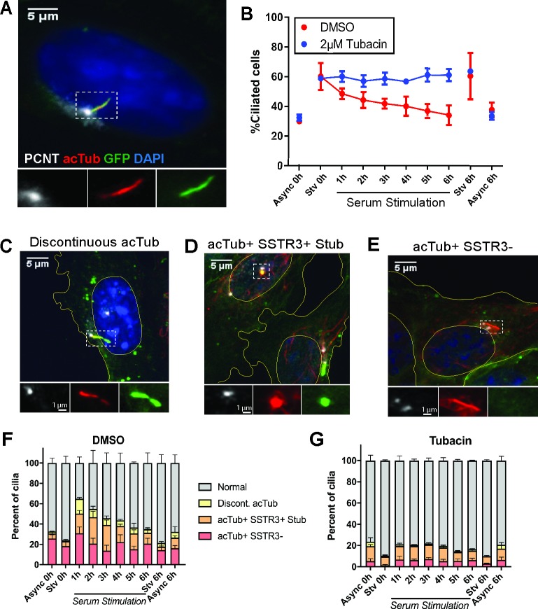
Fig 1. Serum stimulation of IMCD3 cells reveals noncanonical ciliary structures.
IMCD3-SSTR3::GFP cells were serum starved for 24 hrs, followed by stimulation with 10% serum in order to synchronize ciliary loss. A, C–E) Cells were fixed at indicated time points after serum addition and immunostained for PCNT to mark the basal body (white) and acTub to mark the axoneme (red). Nuclei and cell boundaries are outlined in yellow. A) Morphology of a normal, intact cilium in a starved cell. B) The population of ciliated cells quantified over a serum-stimulation time course. Async and serum-Stv controls were included at 0 hrs and 6 hrs. Cells treated in parallel with 2 μM tubacin. C–E) Noncanonical ciliary structures identified in serum-stimulated cell populations. C) Discontinuous acTub staining, in this case accompanied by narrowing of the membrane. D) A ciliary stub, marked by punctate acTub and SSTR3 fluorescence. E) Full-length axoneme marked by acTub, lacking corresponding SSTR3::GFP signal. F–G) Stacked plots of cilia morphologies observed during serum stimulation in (F) DMSO and (G) tubacin-treated cells. Quantifications are based on means of three independent experiments with 150–200 cells analyzed per condition per replicate. Error bars = SEM. Source data can be found in supporting data file S1 Data. acTub, acetylated tubulin; Async, asynchronous; IMCD3; PCNT, pericentrin; SSTR3::GFP, somatostatin receptor 3::green fluorescent protein; Stv, starved.

