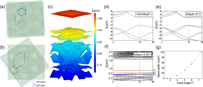
Figure 1.
Atomic and electronic structures of TBBN. (a,b) Schematic illustration of the two possible configurations in twisted bilayer hBN that have the same Moiré pattern. The B/B regions (highlighted with solid blue circle) and the N/N regions (highlighted with dash red circle) are located in different sites in the supercell in configuration α (a), whereas they share the same sites in configuration β (b). (c) Calculated DFT band structure of unrelaxed twisted bilayer hBN at 2.64° in a region of 0.11 × 0.11 1/Å2 around the Γ-point in the supercell Brillouin zone. (d–f) DFT band structures of monolayer (d), normal bilayer without twist (e), and unrelaxed twisted bilayer hBN (f). (g) Band width of the first set of flat bands at the top of the valence bands of unrelaxed TBBN for different twist angles.

