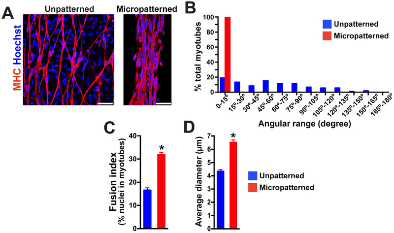
Figure 2. Micropatterning on rigid substrate enhances alignment and fusion of iPSC-derived myotubes.
(A) Representative images of immunocytochemistry labeled myosin heavy chain (MHC) after 2 weeks of terminal differentiation on a patterned or unpatterned rigid substrate. Multiple nuclei were lined up and fused in MHC+ myotubes on micropatterned lanes. In contrast, single fusion was identified for cells cultured on unpatterned geometry. Scale bar = 50 μm. (B) The myotubes on micropatterned lanes (n = 100) were highly-aligned within a 15° angle of lane direction compared to the myotubes on an unpatterned platform (n = 116). (C) Fusion index analysis revealed that more nuclei were fused together in MHC+ myotubes on micropatterned lanes compared to the myotubes on unpatterned platforms. Fifteen and sixteen microscopic fields from micropatterned and unpatterned culture were used for analysis respectively. (D) Micropatterned geometry increased myotube diameter. (n = 100 in micropatterned, n = 116 in unpatterned; *p< 0.01)

