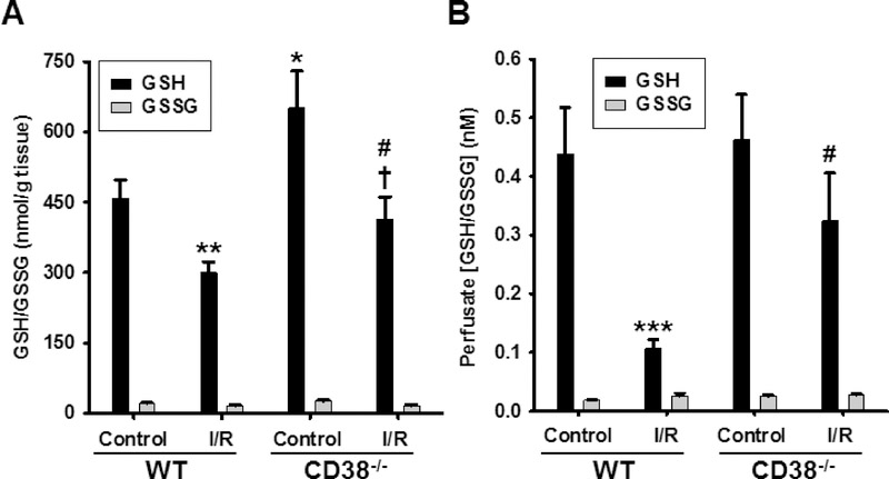
Figure 8. Myocardial and endothelial glutathione levels in WT and CD38−/− hearts undergoing control perfusion or I/R.
HPLC with fluorescence was used to measure GSH-OPA adducts before or after NEM labeling with subsequent reduction by DTT to assess GSH and GSSG, respectively in A) heart tissue, and B) coronary effluent of endothelium-permeabilized hearts. In heart tissue, I/R resulted in significant reduction in GSH levels in both groups with about 35% reduction seen for each. However, baseline GSH levels were significantly higher in CD38−/− compared to WT hearts. Analysis of coronary effluent after endothelial pemeabilization showed marked GSH depletion from the endothelium of WT hearts. In CD38−/− hearts, this depletion was much less severe and did not reach statistical significance, indicating protection of post-ischemic endothelial GSH pools in CD38−/− hearts. *P<0.05, **P<0.01, ***P<0.005 compared to WT Control, †P<0.05 compared to CD38−/− control, #P<0.05 compared to WT I/R. Mean ± SEM; n= 5–8).

