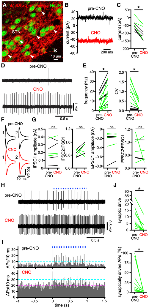
Figure 5. Chemogenetic Activation of hM3D(Gq)-mCherry in STN Neurons Augmented Their Intrinsic Activity.
(A) Confocal micrograph of hM3D(Gq)-mCherry expression (red) in NeuN-immunoreactive STN neurons.
(B and C) Chemogenetic activation of hM3D(Gq)-mCherry with 10 μM of CNO generated a persistent inward current in STN neurons that were voltage clamped at −60 mV in ex vivo brain slices prepared from 6-OHDA- (green) or vehicle- (black) injected mice (B, example from a 6-OHDA-injected mouse; C, population).
(D and E) Chemogenetic activation of hM3D(Gq)-mCherry increased the frequency and regularity of autonomous STN activity ex vivo (D, example from a 6-OHDA-injected mouse; E, population, black and green from vehicle- and 6-OHDA-injected mice, respectively).
(F and G) Pairs of inhibitory postsynaptic currents (iPSCs; upper black and red traces; holding voltage, −60 mV) and excitatory synaptic currents (EPSCs; lower black and red traces; holding voltage, −80 mV) electrically evoked in STN neurons with an interval of 100 ms were unaffected by chemogenetic activation (F, examples from a 6-OHDA-injected mouse; G, population data, black and green from vehicle- and 6-OHDA-injected mice, respectively).
(H–J) The patterning of STN activity by motor cortical inputs optogenetically stimulated at 20 Hz for 1 s (blue) in ex vivo brain slices from 6-OHDA-injected mice was reduced following chemogenetic rescue of intrinsic activity (H, example cell-attached recordings; I, population peristimulus time histogram [magenta line, pre-stimulus mean; cyan line, pre-stimulus mean + 3 SD]; J, population data; data were derived from 5 trials of stimulation per neuron). APs, action potentials.
*p < 0.05. ns, not significant.
See also Table S5.

