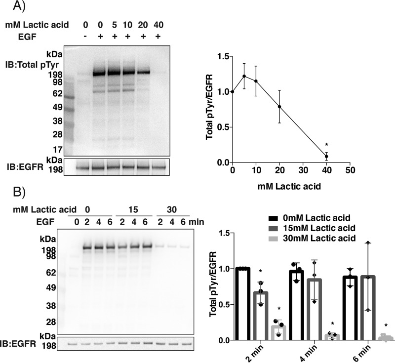
Figure 4.
Treatment of A431 cells with lactic acid prior to EGF ligand stimulation results in less phosphorylation. A, concentration dependence. Serum-starved A431 cells in regular DMEM containing bicarbonate were pretreated for 2 min with 0, 5, 10, 20, and 40 mm lactic acid and subsequently stimulated with EGF (100 ng/ml) for 5 min. Lysates were analyzed for total phosphotyrosine. B, time course. A431 cells were pretreated for 2 min with 0, 15, and 30 mm lactic acid and subsequently stimulated with EGF (100 ng/ml) for 2, 4, and 6 min. Lysates were analyzed for total phosphotyrosine. The right-hand panels in A and B show densitometry analyses as in Fig. 3 with filled circles showing individual results (n = 3; mean ± S.D. (error bars); *, p < 0.05). The membranes in this figure stained with Ponceau for total protein loading are shown in Fig. S4.

