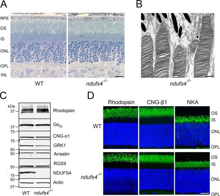
Figure 2.
Normal photoreceptor morphology in ndufs4−/− mice. A, plastic cross-sections of WT (left) and ndufs4−/− (right) retinas at P47. Scale bar, 10 μm. OS, outer segments; IS, inner segments; ONL, outer nuclear layer; OPL, outer plexiform layer; INL, inner nuclear layer. B, electron micrograph of distal outer segments of photoreceptors from a P47 ndufs4−/− retina. Scale bar, 1 μm. C, Western blot analysis of retinal lysates (25 μg loaded per lane) determining expression levels of NDUFS4 protein and of representative proteins relevant to phototransduction in WT and ndufs4−/− retinas. Actin is the loading control. Gαt1, rod transducin; GRK1, rhodopsin kinase; RGS9, regulator of G-protein signaling member 9. D, immunolocalization of rhodopsin, CNG-β1, and the Na+/K+-ATPase (NKA) within rod photoreceptors. Scale bar, 20 μm.

