Figure 4.
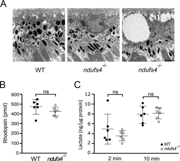
Retinal pigment epithelium morphology and function in ndufs4−/− mice. A, electron micrographs of RPE cells at P47. Although most ndufs4−/− RPE (middle panel) had a morphology similar to WT (left), occasional ndufs4−/− RPE cells (right) were observed to have giant intracellular vacuoles. Scale bar, 2 μm. B, rhodopsin content of isolated WT and ndufs4−/− retinas at P47, as measured by difference spectroscopy. n = 6 retinas per genotype. C, quantification of lactate liberated from isotonic washes of isolated retinas. In separate experiments, WT and ndufs4−/− retinas were washed for 2 or 10 min, and the lactate content was normalized to total retinal protein for each sample. n = 6 retinas per genotype in each experiment. Bars indicate mean ± S.D. ns, not significant.

