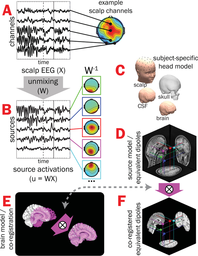
Figure 1.

Subject EEG effective source separation, dipole localization, and inter-subject co-registration. A) Time-varying EEG recorded from a subset of scalp-positioned channels are plotted for a single subject during one correctly-performed trial of the flanker task (stimulus-onset = dashed vertical line, button-press = solid vertical line); note that scalp EEG for a given channel is a spatiotemporal admixture of activities from multiple brain and non-brain sources, leading to multiple “hot spots” in scalp projections. B) Adaptive Mixture Independent Component Analysis (AMICA) was used to “unmix” scalp-recorded channel data (by finding the un-mixing matrix, W) into putative EEG effective “source” signals with maximal temporal independence (only a subset shown here on the left) and associated scalp topographies (middle, W−1). C) Subject-specific head models created from structural MRIs were then used with scalp topographies to estimate the coordinates of each source’s equivalent current dipole (D). E) Finally, alignment parameters mapping a given subject’s brain volume (magenta) onto the MNI template brain (gray) were obtained and applied to dipole coordinates, resulting in all subjects’ source model dipoles having a common neuroanatomical reference (F). X = scalp-recorded EEG (channels-by-time); W = unmixing matrix derived from ICA; u = EEG effective source activations (sources-by-time); W−1 = mixing matrix (component scalp topographies).
