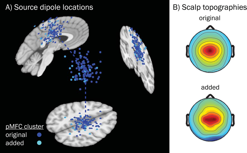
Figure 5.

Equivalent current dipoles and mean scalp topographies for the pMFC source cluster derived by the k-means algorithm and post hoc additions. A) Subject dipoles and their projections onto MNI template space for pMFC are smaller in size than the cluster centroid (located in right mid-cingulate cortex, MNI coordinates: 4, −12, 44), which is larger. Blue subject dipoles depict sources that were included in the original k-means clustering output (n = 94, across 78 subjects); cyan dipoles correspond to sources that were added (for subjects with no dipole in the original cluster, n = 16) subsequently based on nearest distance in clustering measure space. B) Mean scalp topography for sources that were originally included in the k-means obtained pMFC cluster (above), and the topography for those sources which were subsequently added (below). Added subjects did not differ from the original pMFC-clustered subjects in terms of demographics (female vs. male: t[22] = −1.10, p = .284; age: t[21] = −.72, p = .477, Welch’s two-sample t-test) or study variables of interest (error rate [log-transformed]: t[78] = 1.00, p = .319; ADHD symptoms [log-transformed]: t[78] = −1.48, p = .142, MLM linear regression). Note that while some dipole coordinates appear as being located outside of the brain for a given sagittal, coronal, or axial MRI slice (slices chosen based on the centroid coordinates), all dipoles were contained within the boundaries of the template brain as part of their inclusion criteria.
