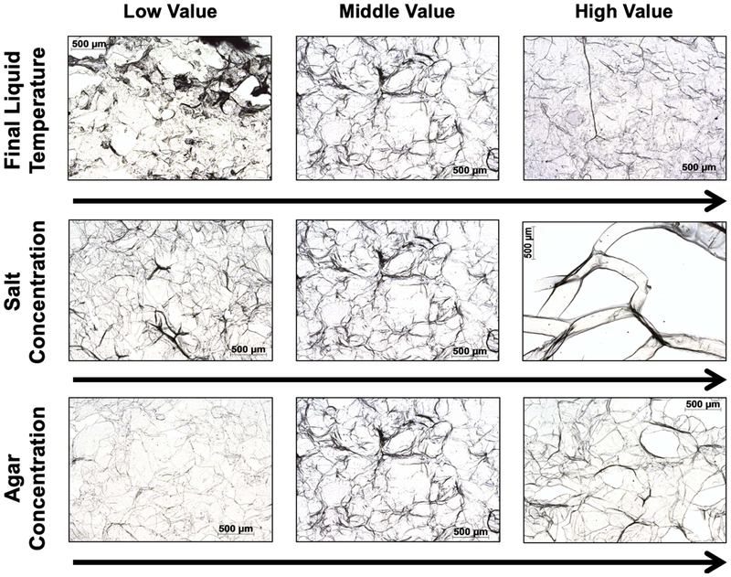
Figure 9.
Microscopy images of agar gel demonstrating microstructural changes that arise in agar phantoms with preparation protocol. Middle value images were created at 90 °C, 0.6%wt salt concentration, and 1.0% agar concentration. The other images were created by holding other parameters and varying only parameter of interest: (Top) Temperature (84, 90, 96 °C); (Middle) salt concentration (0.3%wt, 0.6%wt, 0.9%wt); and (Bottom) agar concentration (0.6%wt, 1.0%wt, 1.4%wt).

