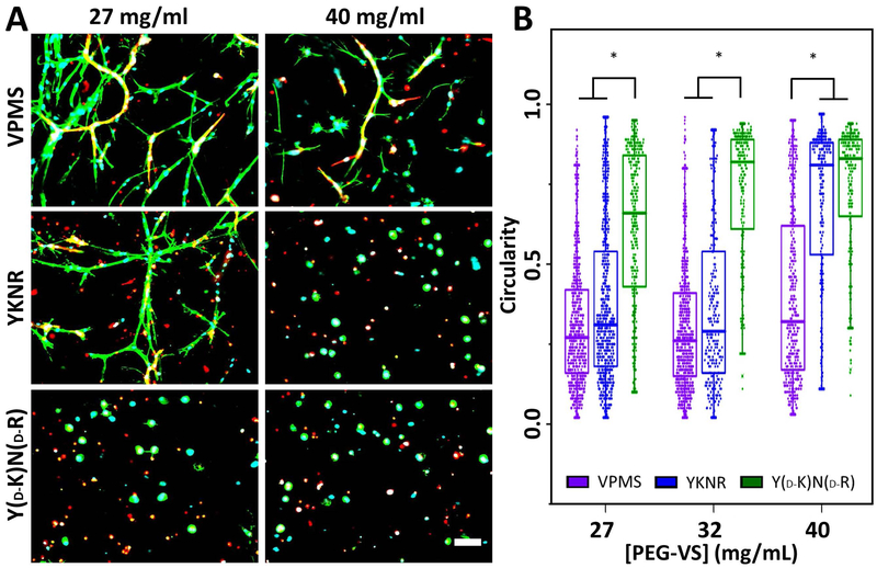
Fig 4:
Reduced crosslinking density allows for fibroblast spreading in YKNR crosslinked PEG-VS hydrogels. ECs and DFs were encapsulated in PEG-VS hydrogels with varying peptide identity and crosslinking density, controlled by initial PEG-VS concentration. UEA (red), phalloidin (green), and DAPI (blue) co-stained images from the centers of the constructs are shown for loose (27 mg/mL PEG-VS) and dense (40 mg/mL) VPMS, YKNR, and YD-KND-R crosslinked hydrogels (A). Fibroblasts are phalloidin positive but UEA negative. Scale bar: 100 μm. Circularity was quantified (see methods) as a measure of cell spreading in loose (27 mg/mL PEG-VS), intermediate (32 mg/mL), and dense (40 mg/mL) crosslinked hydrogels (*: p ≤ 0.003 for comparison shown) (B).

