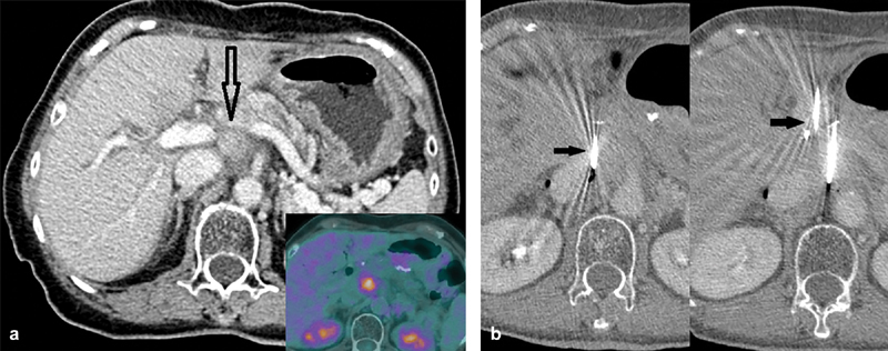
Fig. 1.

Case 1. A 73-year-old woman with recurrence of pancreatic adenocarcinoma post–pancreatico-duodenectomy (Whipple's). ( a ) FDG avid enhancing soft tissue in the surgical bed (inset). Notice the close proximity to the portal vein, inferior vena cava, celiac trunk, and splenic vein (empty black arrow). ( b ) First and second needle placement adjacent to the inferior vena cava and the aorta. Notice the needles placed parallel and ∼1.5 cm apart (arrows). Optimal spacing between IRE probes are 1.5–2.2 cm. Parallel placement is needed to ensure nondistortion of the electrical field, although in practice up to 10 degrees divergence is acceptable.
