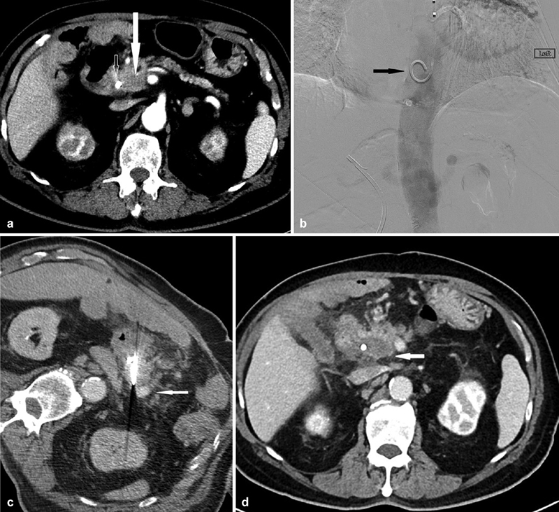
Fig. 4.

A 70-year-old man presenting with a tumor in the head of the pancreas who was medically unfit for pancreatico-duodenectomy. ( a ) Preprocedural CT showing a hypoenhancing mass (closed arrow) in the head of the pancreas surrounding by the superior mesenteric artery, hepatic artery, gastroduodenal artery, as well as the common bile duct with a biliary stent in situ (open arrow). The superior mesenteric vein has been completely obliterated by the mass. ( b ) Placement of a pigtail catheter (arrow) in the descending aorta through a left transradial approach before IRE. This aids in the visualization of the vascular anatomy through 10-mL boluses of 50% contrast during the placement of IRE probes during intermittent CT fluoroscopy. ( c ) Placement of IRE probes. Note the enhancement of the surrounding vasculature (arrow) which would otherwise be difficult to delineate without intermittent contrast boluses from the preplaced pigtailed catheter in the descending aorta. ( d ) CT 3 months postablation showing no evidence of recurrence at the ablation site (arrow) with stability in size.
