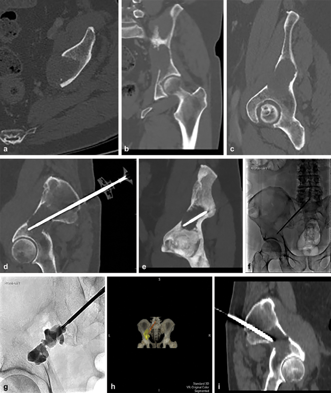
Fig. 4.

( a–c ) CT axial, coronal, and in plane views of left periacetabular lesion within the ilium demonstrating complete cortical erosion of the medial wall of the ilium. ( d, e ) CT of polymethyl methacrylate (PMMA) injection trochar positioned in the lesion following RFA. ( f, g ) Fluoroscopic images showing injection of PMMA. ( h ) 4D CT showing a posterior view of the pelvis, the trochar, and the lesion filled with PMMA. ( i ) Sagittal view of the pelvis demonstrating the course of the screw.
