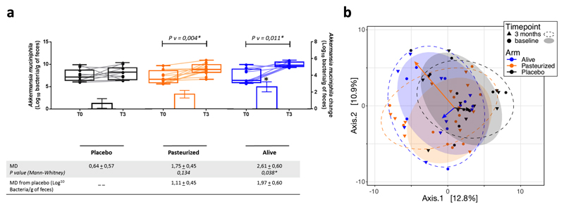
Extended data figure 3. Changes in faecal microbiome.
(a) Akkermansia muciniphila abundance in feces evaluated by Q-PCR. Differential values (MD and MD from placebo) are expressed as mean ± SEM as raw. Bars represent mean change from baseline value per group, with their standard errors. Mann-Whitney tests were performed to compare differential values of both treated groups versus the Placebo group (inter-group changes), according to the distribution. The respective P value are indicated in the table. Lines represent raw values before and after 3 months of supplementation. Distribution of values within each group for each timing is illustrated by a box and whisker. In the box plots, the line in the middle of the box is plotted at the median, the inferior and superior limit of the box correspond to the 25th and the 75th percentiles respectively. Wilcoxon matched-pairs signed ranks tests were performed to verify changes from baseline (intra-group changes), according to the distribution. When the difference is significative, a capped line is marked above the concerned group with the corresponding P value. Kruskal-Wallis analysis were used to compare changes between 0 and 3 months across the 3 groups, according to the distribution. MD, Mean Difference. Placebo, n = 11; Pasteurized, n =12; Alive, n=9. All tests were performed in two-tailed. *P < 0.05.
(b) Visualization of the participants’ faecal microbiota composition at baseline and endpoint of the intervention. Faecal microbiota dissimilarity between samples is represented by principal coordinates analysis (genus-level Aitchison distance), with 6 sample groups - corresponding to the 3 different treatment arms at baseline or at endpoint represented by confidence ellipses (80% confidence interval). Intervention effects are symbolized by colored arrows, with direction and length corresponding to the shift in group centroid coordinates from baseline to endpoint for each treatment arm (re-scaled x4 and re-centered at baseline global centroid). Placebo, n = 11; Pasteurized, n =12; Alive, n=9.

