Figure 1.
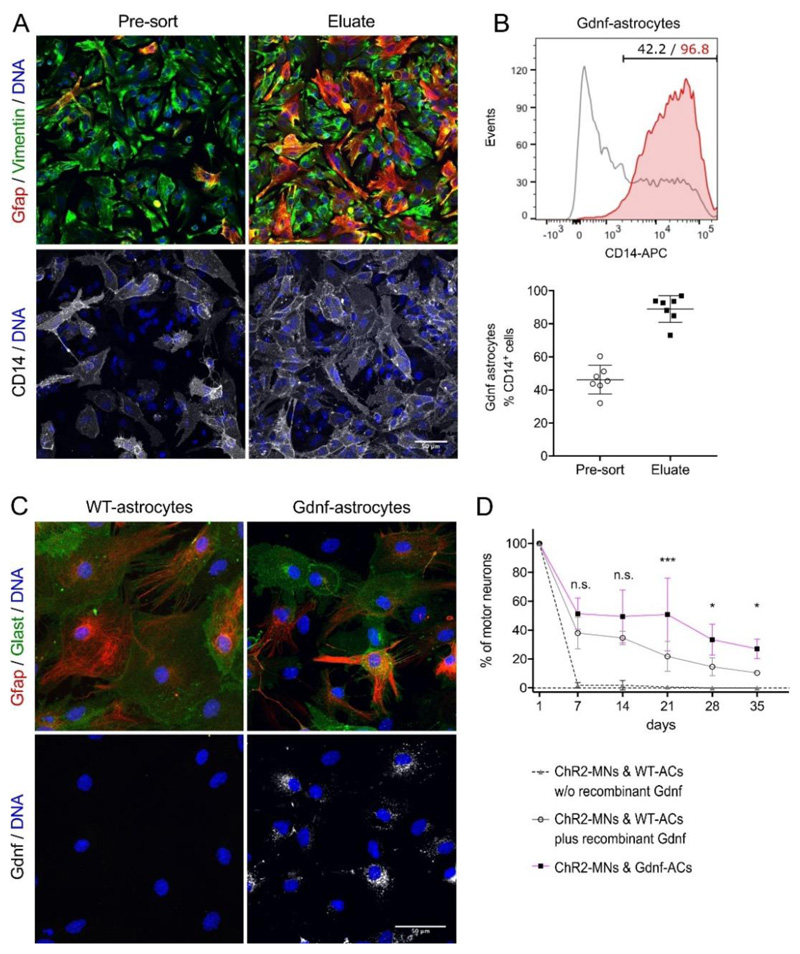
ESC-ACs differentiated from GFAP::CD14/CAG::Gdnf ESCs. (A) Gdnf-AC cultures without and with anti-CD14 enrichment by MACS. Cells were labelled for the AC markers Gfap and Vimentin; scale bar 50μm. (B) Top: Representative flow cytometry analysis of anti-CD14-enriched ACs showing the percentage of ACs in differentiation cultures prior (black) and after (red) MACS. Bottom: AC enrichment was quantified in n=7 independent experiments. Lines indicate mean and SD, individual experiments are represented by points. (C) Gdnf detection in WT and Gdnf-ACs. Cell were labelled for the AC markers Gfap and Glast, and for Gdnf; scale bar 50μm. (D) Long term survival of motor neurons co-cultured with WT-ACs in medium without (dashed line) or with recombinant Gdnf (solid grey line) and Gdnf-ACs (magenta line) in medium without Gdnf (n=3, each experiment in triplicate).
From day 7 onwards all data points of ChR2-MN/WT-AC without recombinant Gdnf are significantly different from both other groups (not shown in the figure, p < 0.0001). Two-way ANOVA, *p<0.05, *** p<0.001, n.s. not significant (p > 0.05). Error bars indicate SD.

