Figure 3.
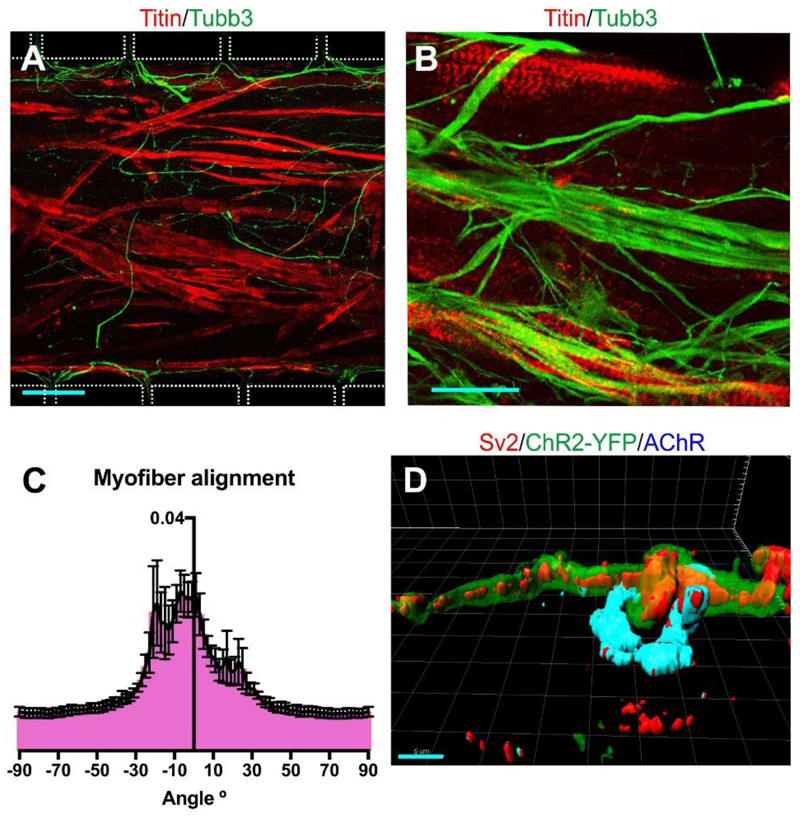
Morphological analysis of myofibres. (A) Fused myofibres in the central compartment at day 9 were identified by Titin labelling; β3-tubulin labels motor axons. The outlines of the compartment boundaries and microchannels are shown as a white dotted line. Scale bar 100 μm (B) Titin staining of the cultured myofibres shows that they have matured and show striations. Scale bar 25 μm. (C) Myofibres preferentially align with the long axis of the central compartment (0° angle), as determined by Fourier analysis (n=6 z-stacks of different devices). Error bars indicate SD. (D) 3D-reconstruction of an in vitro NMJ. The sample was stained with antibodies to the pre-synaptic marker Sv2, YFP, and the postsynaptic marker AChR. Scale bar 5 μm.

